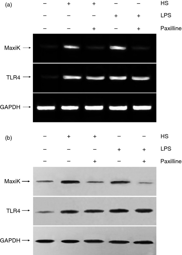
Figure 1.

The mRNA and protein expression of MaxiK and Toll-like receptor 4 (TLR4) in RAW264.7 cells. RAW264.7 cells were maintained in Dulbecco's modified Eagle's medium supplemented with 10% fetal bovine serum at 37° with 5% CO2. Cells were incubated for 6 hr at 37° with heparan sulphate (HS; 1 μg/ml) or lipopolysaccharide (LPS; 10 ng/ml) in the absence and presence of paxilline (2 μg/ml), respectively. (a) To determine the mRNA expression levels of MaxiK and TLR4, total cellular RNA was isolated from the cells and reverse transcribed with primers. After amplification by PCR, the products were separated by electrophoresis in 1% agarose gels containing 0·1 μg/ml ethidium bromide and visualized under ultraviolet light. (b) To detect the protein expression of MaxiK and TLR4, whole cell lysate was separated on SDS–polyacrylamide gels followed by transfer to a PVDF membrane. The membrane was incubated with blocking solution for more than 1 hr at room temperature and subsequently incubated with anti-MaxiK or TLR4 antibody (1 : 1000 dilutions) overnight at 4°, respectively. After 1-hr incubation at room temperature with horseradish peroxidase-conjugated secondary antibody (1 : 2000 dilution), the protein bands were visualized by the chemiluminescent detection system. Results of one out two experiments with similar results are shown.
