Figure 3.
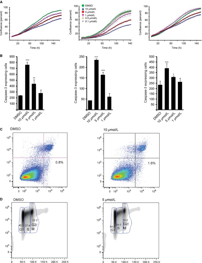
JW74 treatment inhibits osteosarcoma (OS) growth. (A) The proliferative capacity of KPD, U2OS and SaOS-2 was inhibited following treatment with JW74 (1–10 μmol/L). Cell densities were measured by IncuCyte live cell imaging. DMSO was included as control. (B) The number of Caspase-3-expressing cells per well, following 52 h exposure to drug was determined using the IncuCyte live cell imaging system. Caspase-3 activity was significantly increased in a dose-dependent manner (*P = 0.014; **P = 0.008; ***P < 0.001). Cells were treated as described in (A), including Cell player reagent in the culturing medium, which renders cells expressing increased levels Caspase-3 fluorescent. (C) The percentage of apoptotic U2OS cells increased from 0.8% (DMSO) to 1.6% (10 μmol/L JW74) following 72 h drug treatment was determined by Alexa-488 Annexin V binding (x-axis). Propidium iodide (PI) was included as a marker of necrotic cells (y-axis). The analysis was performed by flow cytometry. A representative experiment is shown (D) JW74 treatment leads to accumulation of U2OS cells in G1 phase. The cells were treated with 0.1% DMSO (control) or 5 μmol/L JW74 for 72 h and subsequently labeled with Hoechst (x-axis) and stained with proliferation marker Ki67 (y-axis). The number of cells in each cell cycle phase was determined by flow cytometry. A representative experiment is shown.

