
Fig 3.
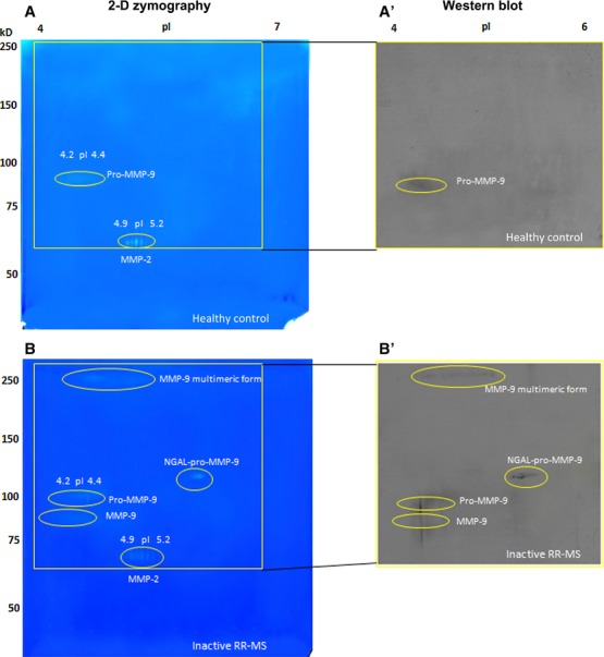
2-DZ analysis and identification of MMP-9 by Western blot. Left panels (A and B): 2-D zymography (2-DZ) of serum samples from healthy control (A) and from an inactive RR-MS patient not subjected to therapy (same patient as in Fig. 2; B). For 2-DZ, aliquots of 35 μl of serum were resuspended in the rehydration solution and subjected to isoelectrofocusing (IEF; 1st dimension) on IPG Dry-Strips of 13 cm in a linear pH gradient of 4–7. After IEF, IPG strips were equilibrated and then applied for the 2nd dimension in a 8.5% (w/v) polyacrylamide gel copolymerized with 0.1% (w/v) gelatin. The isoforms and charge variants of MMP-2 and MMP-9 appear as clear spots of digestion on the dark background of the gel. Right panels (A' and B') represent Western blot analysis of the same sera shown in A and B. Aliquots of 35 μl of serum samples (instead of the usual 20 μl) were subjected to 2D electrophoresis (2-DE) by using 8.5% (w/v) polyacrylamide gels without gelatin. After transfer of the proteins, the nitrocellulose membranes were incubated with an anti MMP-9 (Ab-8) Mouse mAb (IA5) at concentration of 2.66 μg/ml.