
Fig. 2.
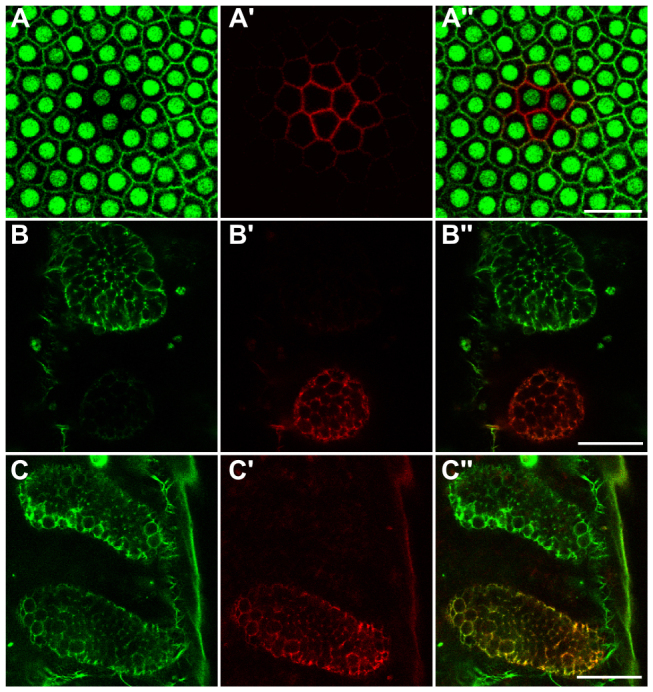
Fluorescence labeling of Tribolium embryos with photoconvertible ABP-tdEosFP. (A-A′) Optical sections through the blastoderm of a transgenic embryo ubiquitously expressing nuclear GFP (Sarrazin et al., 2012), which was injected with ABP-tdEosFP mRNA and photoconverted in the central region. (A) nGFP and unconverted ABP-tdEosFP fluorescence detected at similar levels in the green channel, (A′) photoconverted ABP-tdEosFP fluorescence detected in the red channel and (A′) overlay of the two channels. (B-B′) Optical sections through developing limb-buds of an embryo 26 hours after injection with ABP-tdEosFP mRNA. Most of the protein in the bottom limb-bud has been photoconverted. (B) Green channel showing unconverted ABP-tdEosFP, (B′) red channel showing converted ABP-tdEosFP, and (B′) overlay of the two channels. (C-C′) Optical sections through the same limbs shown in B-B′ 22 hours later. Both green unconverted and red converted ABP-tdEosFP are detected in the bottom photoconverted limb. Anterior is towards the top in all panels, and ventral midline is towards the left in B-C′. Scale bars: 25 μm.