
Fig. 6.
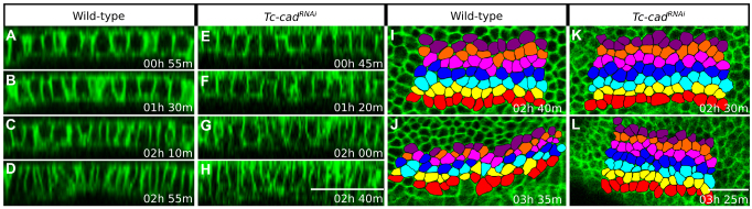
Cell behaviors during Tribolium germband condensation in wild-type and Tc-cadRNAi embryos. (A-H) Progressive contraction of embryonic cells observed both in (A-D) wild-type and (E-H) Tc-cadRNAi embryo at four time-points spanning, from top to bottom, stages 1 to 3. Panels show transverse sections of GAP43-YFP-labeled wild-type and Tc-cadRNAi embryos, also shown in Fig. 4A-E and Fig. 4K-O, respectively. Panels are timed against the onset of stage 1 and apical is towards the top. (I-L) Cell intercalation in wild-type embryo (I,J) and absence thereof in Tc-cadRNAi embryo (K,L). Panels show ventrolateral views of GAP43-YFP-labeled embryos at two time-points during stages 3 (top) and 4 (bottom). All panels are arranged with anterior towards the left and show average intensity projections of 6 μm substacks covering the anterior left side of the condensing germbands. Abutting rows of ectodermal cells, highlighted with different colors, were tracked after their 13th division. (I,J) Cells in each row become separated by ventral or dorsal neighboring cells in the wild type. This cell intercalation narrows the highlighted cluster of cells in the dorsal-ventral axis and lengthens it in the anterior-posterior axis. Many cells also appear to elongate along the anterior-posterior axis. (K,L) Rows of cells remain contiguous as no cell intercalation takes place after Tc-cad RNAi. Under both wild-type and RNAi conditions, the highlighted clusters of cells shrink between the two time-points as a result of cell contraction to about 75% and 73% of their original size, respectively. Scale bars: 50 μm.