
Fig. 7.
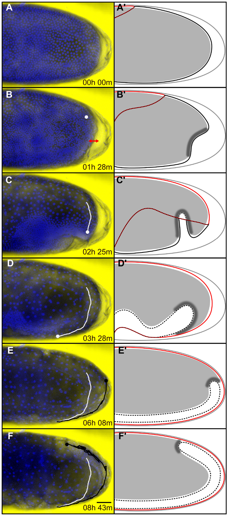
Posterior yolksac dynamics relative to Tribolium germband condensation and elongation. Time series of posterior yolksac deformation in (A-F) an H2B-RFP-labeled embryo and (A′-F′) corresponding schematic representations. (A-F) Average intensity projections of the posterior half of the same embryo timed against the first time-point shown in A. At each time-point, the DIC image of the embryo was overlaid with the corresponding H2B-RFP signal shown in blue. In B-F, the dot indicates the position of the leading edge of the posterior yolk-fold. The white track marks the early ventral extension of the yolk-fold and the black track marks its dorsal retraction up to the corresponding time-point. In A′-F′, the vitelline membrane is shown with a gray line, the serosa outline with a red line, the amnion/serosa boundary with a dotted red/black line, the condensing and internalized elongating embryonic rudiment with solid and broken black lines, respectively, the yolksac with filled gray, and the presumptive posterior yolksac-germband attachment by darker shading. (A,A′) Uniform blastoderm stage. (B,B′) Differentiated blastoderm stage showing blastoderm and yolk depression at the posterior pole (double-headed red arrow). (C,C′) The posterior dorsal wedge of yolk extends ventrally during posterior amniotic fold formation. There is concurrent amnion involution during yolk-fold extension. (D,D′) The yolk-fold completes its ventral extension when posterior amnion involution stops and serosa window forms. (E,E′) Yolk-fold retraction is tightly coupled to posterior germband extension. The yolk levels out posteriorly when the translucent germband reaches the posterior pole. (F,F′) Yolksac displacement continues dorsally as the translucent germband extends around the posterior pole. Lateral views, anterior towards the left and dorsal towards the top. Scale bar: 50 μm.