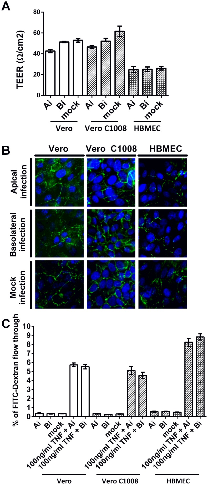
Figure 3. Cell monolayer integrity post CHIKV infection at an MOI of 10.
(A) TEER measurements of Vero, Vero C1008 and HBMEC cell monolayers were taken at 24 h.p.i.. The TEER measurements post-apical and basolateral infection were comparable to that of mock-infected cells. Vertical bars represent one standard deviation from the mean of three readings. (B) Immunofluorescence assays demonstrated the expression of ZO-1 tight junction proteins (green) in apically-infected and basolaterally-infected Vero, Vero C1008 and HBMEC cells at 24 h.p.i.. The expression of ZO-1 proteins in infected cells is comparable to that of mock-infected cells. (C) The FITC-dextran permeability assays demonstrated that the integrity of Vero and Vero C1008 cell monolayers remained intact at 24 h.p.i., where the permeability of the cell monolayers to FITC-dextran remained low as compared to the TNF-treated cells.

