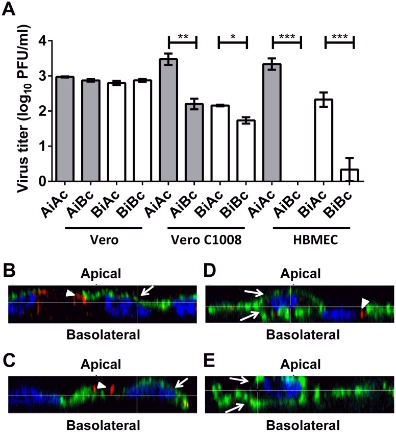
Figure 5. Polarized release of CHIKV at apical plasma membrane domain.
(A) Infectious virus titer of supernatants collected from the apical and basolateral chambers at 24 h.p.i. of non-polarized Vero, polarized Vero C1008 and polarized HBMEC cells at an MOI of 10 were quantified by viral plaque assays. Release of CHIKV is bi-directional in non-polarized Vero cells but occurs preferentially at the apical domain of polarized Vero C1008 and HBMEC cells. Two-tailed Student's t-test: * p<0.05, ** p<0.005, *** p<0.001. Vertical bars represent one standard deviation from the mean of three readings. (B) Apically-infected Vero C1008, (C) basolaterally-infected Vero C1008, (D) apically-infected Vero and (E) basolaterally-infected Vero cells were co-labeled with antibodies against CHIKV E2 glycoprotein (green, arrows) and ZO-1 (red, arrowheads). Cell nuclei were stained with DAPI (blue). Z-stacked images show the polarized release of CHIKV towards the apical plasma membrane of Vero C1008 upon apical and basolateral infection. Release of CHIKV is bidirectional in non-polarized Vero cells.

