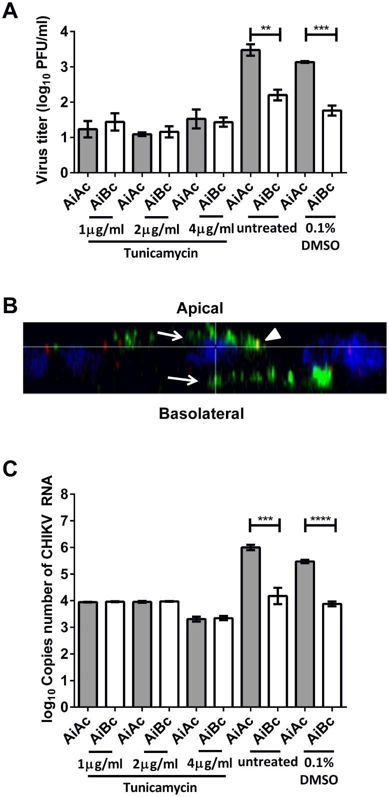
Figure 7. Release of CHIKV upon drug treatment to elucidate viral factors involved in apical sorting of CHIKV.
(A) The infectious virus titers were similar in the apical and basolateral chambers upon treatment with tunicamycin. Vertical bars represent one standard deviation from the mean of three readings. (B) Apically-infected Vero C1008 cells were treated with tunicamycin and co-labeled with antibodies against CHIKV E2 glycoprotein (green, arrows) and ZO-1 tight junction proteins apical markers (red, arrowheads). Cell nuclei were stained with DAPI (blue). Z-stacked images show the bidirectional release of CHIKV upon treatment with tunicamycin. (C) The copies number of CHIKV RNA was higher in the apical chamber than in the basolateral chamber upon apical infection with CHIKV. Upon tunicamycin treatment, the copies number of CHIKV RNA was similar in the apical and basolateral chambers.

