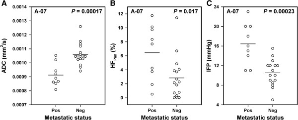
Figure 5.
DW-MRI, hypoxia, IFP, and metastasis data for A-07 melanoma xenografts showing that metastatic tumors had lower ADC, higher HFPim, and higher IFP than nonmetastatic tumors. Median ADC (A), HFPim(B), and IFP (C) for metastatic and nonmetastatic tumors. Points represent single tumors. Horizontal bars indicate mean values.

