
Figure 1.
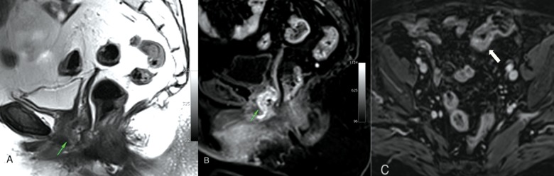
A solid nodular lesion of the anterior vaginal wall appears faintly hyperintense on TSE-T2 sagittal plane (A) and hypervascularized on T1-VIBE contrast enhanced sagittal plane (B). T1-VIBE contrast enhanced axial plane (C) shows a stenosing thickening of the sigmoid wall.