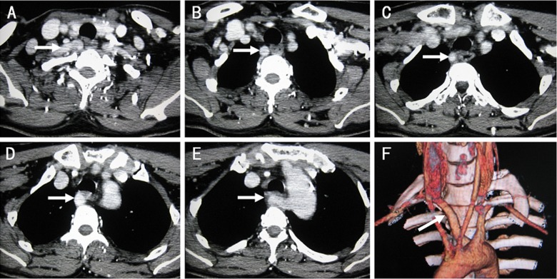
Figure 2.
Contrast-enhanced CT scans of the neck and upper mediastinum in a patient with “arteria lusoria”. From image (A–E), the right subclavian artery (white arrow) originates from the left part of the aortic arch, crossed the mediastinum behind the esophagus, the brachiocephalic artery is absent. Image (F) shows the three-dimensional reconstruction CT scan of “arteria lusoria” (white arrow).

