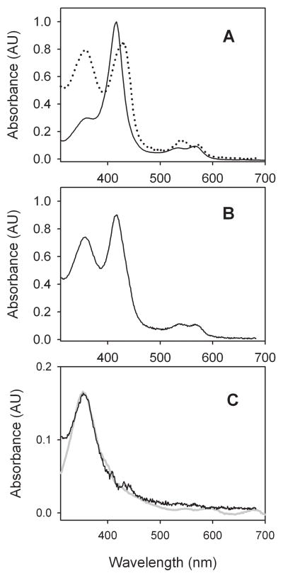
Figure 1.
Spectra from CYP119 in 100 mM phosphate buffer (pH 7) solutions containing 50% glycerol. (A) Ferric enzyme (solid line) and Compound II (dotted line) from PN oxidation at −10 °C. (B) First-formed spectrum following irradiation of Compound II at ca. −40 °C. (C) Residual spectrum after subtraction of ferric enzyme and Compound II spectra from the first-formed spectrum in B (black line) and spectrum of CYP119 Compound I obtained by deconvolution of the time-resolved spectra from reaction of ferric CYP119 with MCPBA (grey line).

