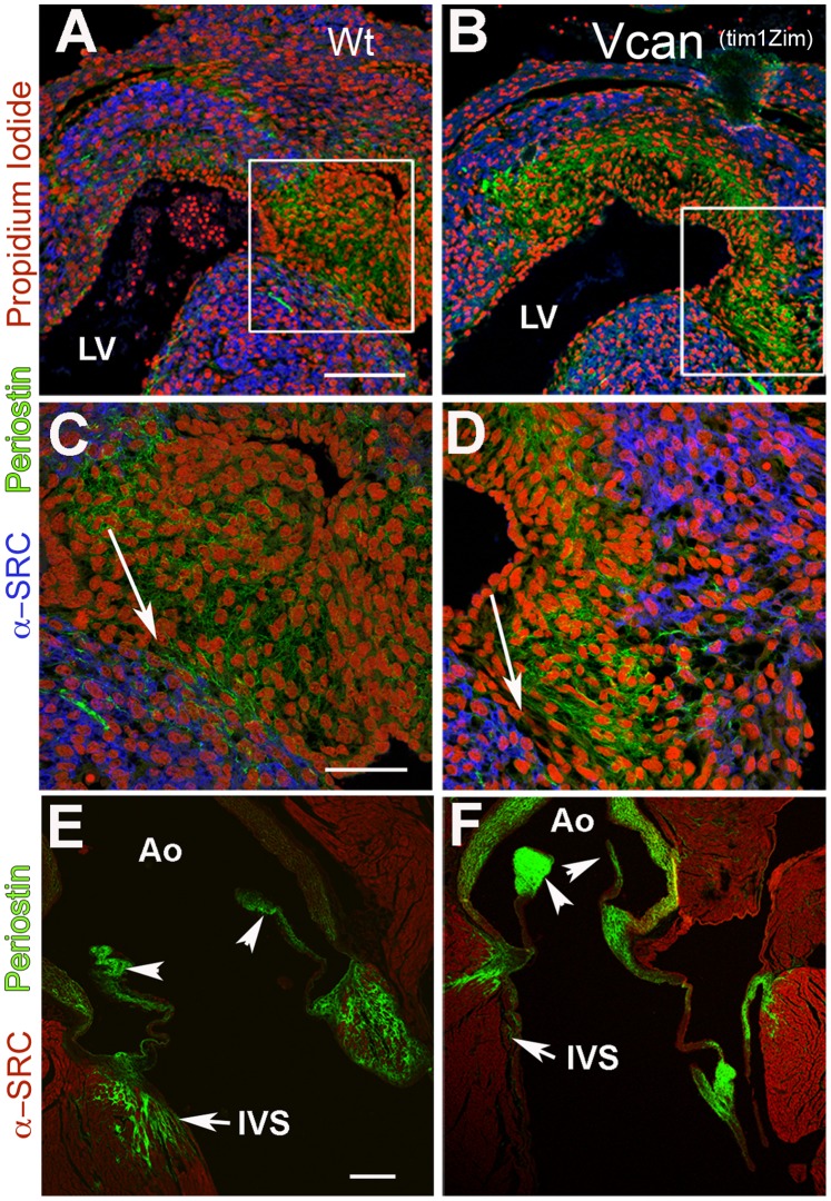
Figure 10. Comparison of periostin in the cushions and valves of Vcan (tm1Zim) embryonic and adult hearts.
Comparisons of periostin expression in wild-type E13.5 pc hearts (panels A, C) and Vcan (tm1Zim) E13.5 pc hearts (panels B,D) by confocal immunofluorescent localization. Periostin (green) expression in the Vcan (tm1Zim) hearts appeared more compact and less widely distributed throughout the cushions than in the wild-type. Boxes in panels A,B denote magnified areas shown in C,D. Periostin staining showed a sharper boundary between the apex of the interventricular septum (blue; α-sarcomeric myosin) and the adjacent cushion mesenchyme in the Vcan (tm1Zim) (D; arrow) as compared to that observed in wild-types (C; arrows). In adult hearts, periostin expression in the Vcan (tm1Zim) (F) appeared more intense in the aorta (Ao) walls and valve leaflets, however very little or no localization of periostin was observed in the interstitial tissue of the muscular interventricular septum (IVS) as compared to wild-type (E). In the Vcan (tm1Zim) heart, periostin appeared to localize only in zones adjacent to the myocardium (F;IVS), but did not show an interspersed pattern of expression between individual myocytes as seen in the wild-type IVS and aortic mitral continuity(E;IVS). Panels A & B same magnification bar = 150 µm; panels C & D same magnification bar = 50 µm; panels E & F same magnification bar = 200 µm.

