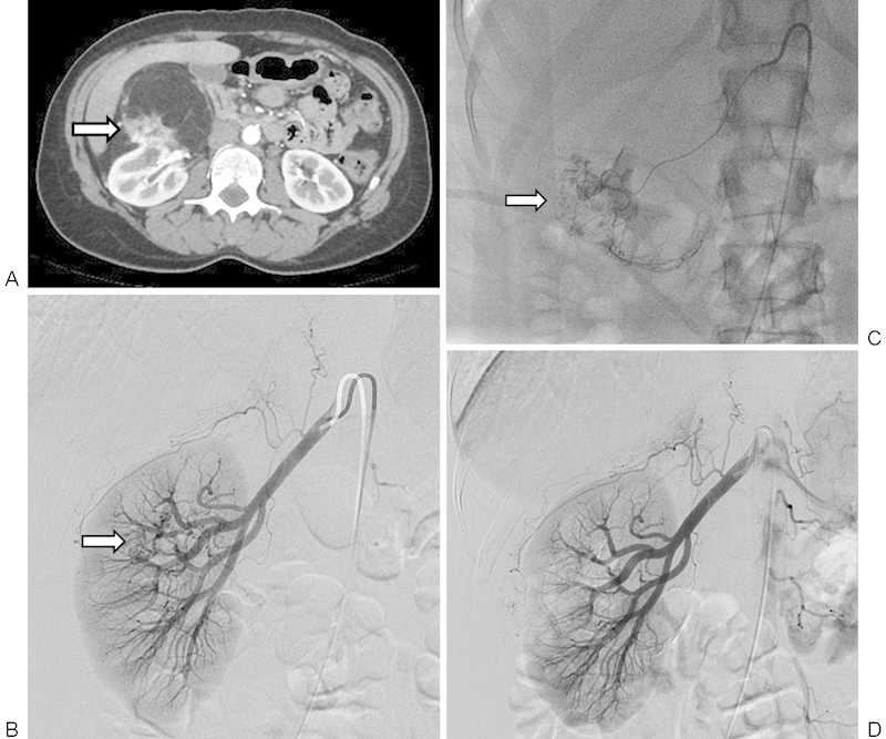
Figure 2.

A 45-year-old woman with incidental 9.4 cm right renal angiomyolipoma that underwent renal arterial embolization. (A) Axial contrast-enhanced computed tomography image of angiomyolipoma demonstrating predominantly fatty components, vascular components (arrow), and origin of the mass from the kidney. (B) Selective right renal arteriogram demonstrating a small area of heterogeneity and hypervascularity overlying the midpole region of the right kidney (arrow). (C) Microcatheter-mediated embolization of the hypervascular aspects of the mass using tris-acryl gelatin microspheres to stasis (arrow). (D) Postembolization selective right renal arteriogram demonstrating complete embolization of the right AML.
