
Figure 4.
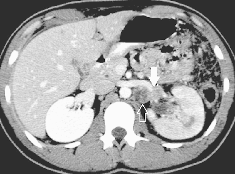
Axial contrast-enhanced computed tomography of a left medullary renal cell carcinoma in a patient with sickle trait. The tumor is multifocal, with invasion of the main left renal vein (white arrow) and extension outside Gerota fascia (hollow arrow) indicating a T stage of T4.