
Figure 2.
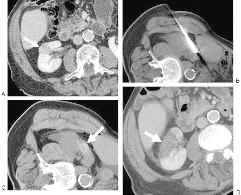
(A) Contrast-enhanced CT of an 83-year-old man shows a 2.3-cm RCC in the interpolar region of the right kidney that extends toward the renal sinus fat (white arrow). The patient was referred for RF because of an overall poor functional status. (B) Noncontrast CT with the patient right-side/ipsilateral-side up demonstrating a lateral approach with the RF probe within the lesion. (C) Noncontrast postablation CT demonstrates areas of increased attenuation (white arrow) in the ablation zone, consistent with blood products, in addition to expected postprocedural changes adjacent to the kidney. This is a common postablation appearance. (D) Contrast-enhanced CT performed approximately 1 month after ablation shows no evidence of enhancement in the ablation zone (white arrow), consistent with a completely treated lesion. CT, computed tomography; RCC, renal cell carcinoma; RF, radiofrequency.