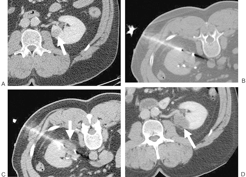
Figure 3.

(A) Contrast-enhanced CT of a 55-year-old man shows a 1.8-cm RCC in the lower pole of the left kidney (white arrow). The patient was referred for cryoablation of the left renal lesion because of prior right nephrectomy for RCC. (B) Noncontrast CT with the patient in prone position demonstrating a posterolateral approach with the cryoprobe within the lesion. (C) Noncontrast CT demonstrates the low-attenuation “iceball” in the ablation zone after a 10-minute freeze (white arrow). (D) Contrast-enhanced CT performed approximately 1 month after ablation shows no evidence of enhancement in the ablation zone (white arrow), consistent with a completely treated lesion. CT, computed tomography; RCC, renal cell carcinoma; RF, radiofrequency.
