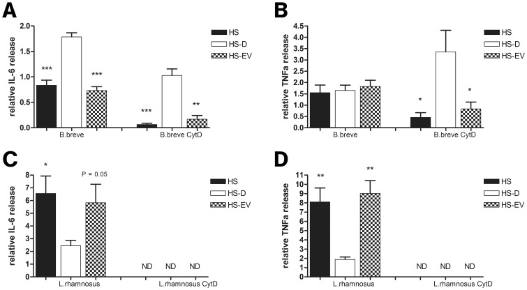
Figure 9. EVs differentially modulate bacterial induced DC cytokine release.
2×10⁵ DCs were co-incubated with 2×10⁶ B. breve NutRes 200 or L. rhamnosus NutRes 1 at 37°C in medium, HS, HS-D or HS-EVs. After 16H, supernatants were collected and analyzed for IL-6 and TNFα release. In another set of similar experiments, DCs were first pretreated with 10 µg/ml cytochalasine D, blocking bacterial phagocytosis. Relative cytokine levels were calculated according to the ratio between responses at serum-free medium and serum fraction supplemented medium. (A) HS and HS-EVs significantly inhibit B. Breve NutRes 200 induced DC IL-6 release compared to HS-D (***P<0.001)(**P<0.01). TNFα release was not affected but upon blocking phagocytosis a significant different TNFα release between HS, HS-EVs and HS-D could be measured (B) (*P<0.05). L. rhamnosus NutRes 1 stimulated DCs release significantly more IL-6 (C) and TNFα (D) in the presence of HS or HS-EVs compared to HS-D. Blocking L. rhamnosus NutRes 1 phagocytosis inhibited DC IL-6 and TNFα release below detection level (ND). Data are represented as mean ± SEM n = 4.

