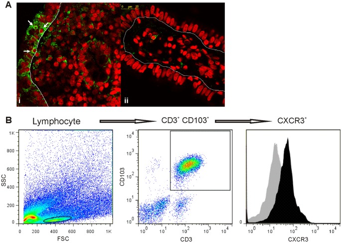
Figure 8. Infiltration of CXCR3+ cells in the intraepithelial compartment.
a. Confocal immunofluorescence for CXCR3 was performed in sections of duodenal biopsies. (i) Intraepithelial lymphocyte CXCR3+ cells in untreated CD patients are indicated by arrows. (ii). CXCR3+ cells were rarely observed in the intraepithelial compartment in non-CD controls. The epithelium is delimited by a thin line. CXCR3 is shown in green, and nuclei are shown in red. (Magnifications, 630× (i) and 1071× (ii)). b. Representative flow cytometric analysis from the epithelial compartment of a duodenal sample of an untreated CD patient showing IELs (CD3+ CD103+) that express CXCR3.

