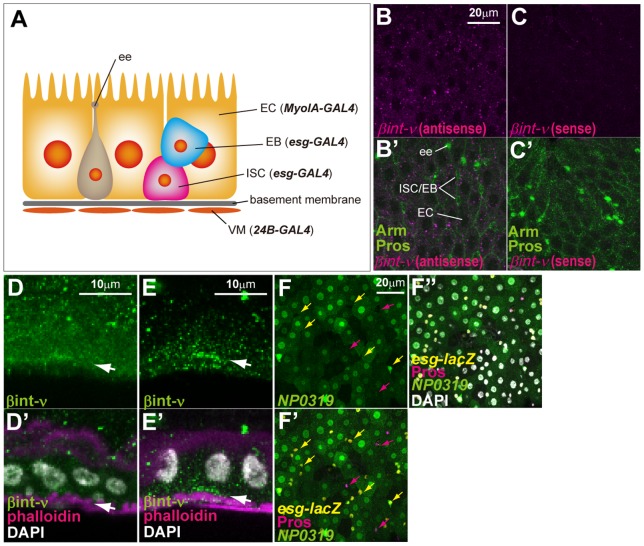
Figure 1. βint-ν is expressed in the adult midgut epithelium.
(A) The diagram of the adult midgut composed of ISCs, EBs, ECs, ees, and VMs. The cell-type specific GAL4 drivers, esg-GAL4, MyoIA-GAL4, and 24B-GAL4, used in this study are shown in parentheses. (B–C) Transcripts of βint-ν were detected with an antisense probe (magenta in B and B’) for βint-ν mRNA but not with a sense probe (magenta in C and C’) for it. “Pros” and “Arm” respectively indicate the ee nuclei and the outline of cells (green in B’ and C’). Small cells without Pros are ISC/EB. (D–E) βint-ν protein was detected with anti-βint-ν antibodies (green, D [30], E [24]). Basal visceral muscles were strongly marked with phalloidin staining (magenta in D’ and E’). Arrows indicate the distribution of βint-ν protein at the basal side. (F-F”) Expression of NP0319 (a βint-ν-GAL4, green), monitored with UAS-GFP, was detected in polyploid ECs and a subset of esg-lacZ-positive cells (yellow) but not the other esg-lacZ- and Pros-positive cells (magenta). Yellow and magenta arrows indicate examples of esg-lacZ- and Pros-positive cells. Nuclei were stained with DAPI (white in D’, E’, and F”).

