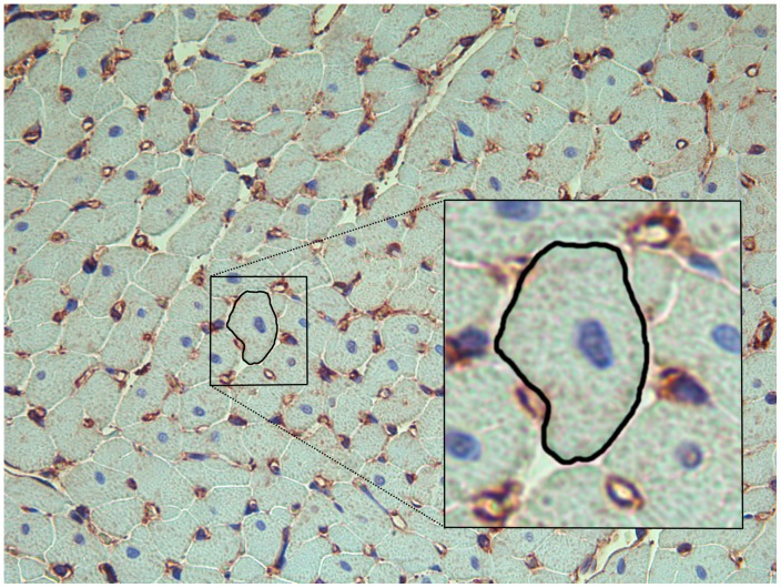
Figure 1. Light microscopy of heart tissue.
Histological section of heart tissue (200x) stained with toluidine blue to enhance the contour of the cardiomyocytes for measuring their circumference and immunohistochemistry using non-muscular β-actin to stain capillaries (brown). One cardiomyocyte cut in short axis and containing a nucleus (blue) is outlined and magnified (box) to show how cardiomyocyte circumference was measured (in arbitrary units).

