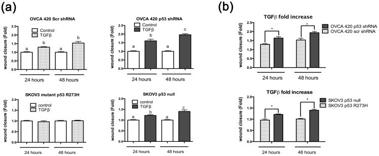
Figure 4. TGFβ induces migration in p53 null cells in comparison to p53 wild-type or mutant cells.
(a) Wound healing assays were performed on SKOV3 and OVCA 420 stable cell lines. Cell monolayers were scratched and treated with or without TGFβ at 20 ng/mL for 48 hours. Wound closure was measured as a fold increase or decrease compared to no treatment control. Paired t-test was used with a p≤0.05. (b) Comparison of the fold increase of TGFβ samples from 5(a). Unpaired t-test was used to analyze significance. Significance is represented by * and signifies a statistical difference between cell lines. Data represented as mean ± SEM, *p≤0.05.

