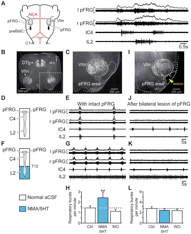
Figure 6. Bilateral lesion of the pFRG suppresses the locomotion-induced accelerated respiration.
A, left, Representation of the brainstem–spinal cord preparation with medullary recording sites. right, Averaged integrated (6 sweeps) and raw respiratory burst activity recorded from the left (l) and right (r) pFRG and from spinal (cervical C4 and lumbar L2) ventral roots. B, C, I, Transverse acetylcholinesterase-stained sections as histological control in intact (B, C) and lesioned (I) brainstem preparations. The position of the lesion ventral to the facial motor nucleus corresponds to the location of the pFRG (I). D, F, Schematics of experimental setup. E, J, Spontaneous respiratory activity recorded bilaterally from the pFRG and homolaterally from cervical (C4) and lumbar (L2) ventral roots under control conditions in intact preparation (E) and after the bilateral lesion of the pFRG (J). G, K, Respiratory effects of lumbar cord activation with NMA/5HT in intact (G) and pFRG-lesioned (K) brainstem-spinal cord preparations. H, L, Bar charts showing variation in the respiratory burst frequency (Resp.; mean ± SEM) under control conditions (open bar, left), during pharmacological activation of lumbar locomotor CPG (blue bar), and after wash-out (open bar, right) in intact (H, n = 7 preparations) and pFRG-lesioned (L, n = 7 preparations) preparations. Recordings shown in E, G, J and K are from the same preparation. VIIn, facial motor nucleus; Ctrl, control conditions; DTg, dorsal tegmental nucleus; D, dorsal; M, medial; WO, wash-out. **P<0.01 (repeated measures ANOVA, Tukey's post hoc tests).

