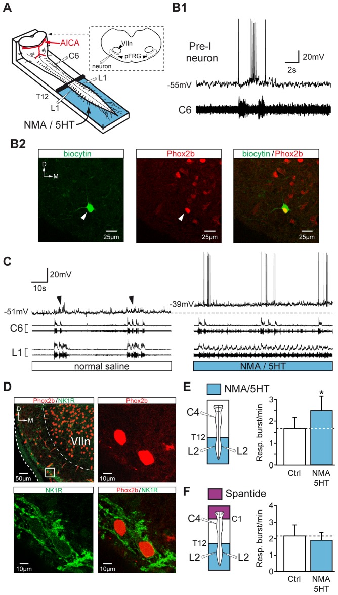
Figure 7. Depolarization of pFRG neurons during pharmacologically induced locomotion in the lumbar spinal cord: involvement of an SP pathway.
A, Schematic representation of the preparation. The brainstem was transected rostrally to the AICA to allow direct access to pFRG neurons for patch-clamp recording. B1, Simultaneous whole-cell patch-clamp recording of a pFRG/Pre-I neuron and raw activity of the cervical (C6, inspiratory-like) ventral root under control conditions. B2, Photomicrographs (z-stack of 3 images, 1.8 µm in total thickness) of immunolabeling for Phox2B (middle, red) in a biocytin-filled neuron (left, green). The white arrowhead indicates the neuron recorded in B1, and the merged image (right) confirms that the double-labeled (yellow) cell was a functionally identified Pre-I neuron. The dashed line shows the ventrolateral edge of the medulla. C, Simultaneous whole-cell patch-clamp recording of a pFRG/Pre-I neuron (black arrowhead indicating spontaneous rhythmic respiratory depolarization) and integrated and raw activity of cervical (C6) and lumbar (L1) ventral roots under control saline conditions (left) and during NMA/5HT application to lumbar region (right). The dashed line indicates the resting membrane potential level under control conditions. D, Left, top, Distribution of NK1R (green) immunoreactivity and Phox2B-positive cells (red) in the ventrolateral part of the medulla (this photomicrograph corresponds to a z-stack of 18 images, 18 µm in total thickness). The inset (white rectangle) shows the ventrolateral aspect of the VIIn at a higher magnification. Note that cells located in the area corresponding to the pFRG networks are immunopositive for Phox2b (right, top, red; z-stack of 3 images, 2.4 µm in total thickness) and for NK1R (left, bottom, green), as confirmed by the merged image (right, bottom). E, F, left, Schematics of experimental procedures. right, Bar charts showing variation in the respiratory burst frequency (Resp.; mean ± SEM) under control saline conditions (open bar) and during NMA/5HT application to the lumbar spinal cord (blue bar) during perfusion of the brainstem with normal saline (E, n = 7 preparations) or with a medium containing the SP antagonist spantide (F, n = 7 preparations). Ctrl, control conditions; D, dorsal; M, medial. *P<0.05 (Student's paired t test).

