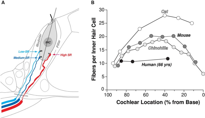
Figure 1.
Innervation of the IHCs by terminals of the cochlear nerve. (A) Schematic illustrating the spatial separation of the synaptic contacts of high- (SR > about 18 spikes/s) vs. medium- and low-SR fibers on the pillar vs. modiolar sides of the IHCs, respectively. (B) Counts of cochlear nerve terminals per IHCs as a function of cochlear location from four mammalian species: cat (Liberman et al., 1990), mouse (Maison et al., 2013), chinchilla (Bohne et al., 1982) and human (Nadol, 1983).

