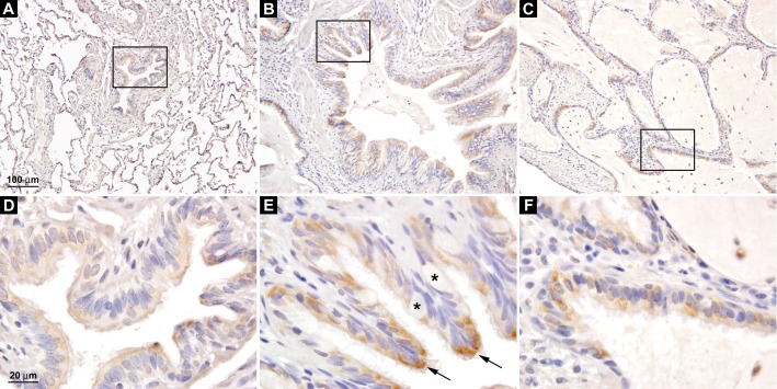
Figure 3.
Expression of HS6ST2S in ciliated bronchial epithelial cells. (A) HS6ST2S immunostaining in the normal lung. Boxed area is shown in D. (B and C) HS6ST2S immunostaining in IPF lungs. Boxed areas are shown in E and F. HS6ST2S was expressed in ciliated bronchial epithelial cells (arrow in E), whereas the mucus-producing goblet cells were negative for HS6ST2S (asterisks). A–C are at the same magnification, and D–F are at the same magnification. Images shown are representative of data from five normal and seven IPF lungs.

