Figure 2.
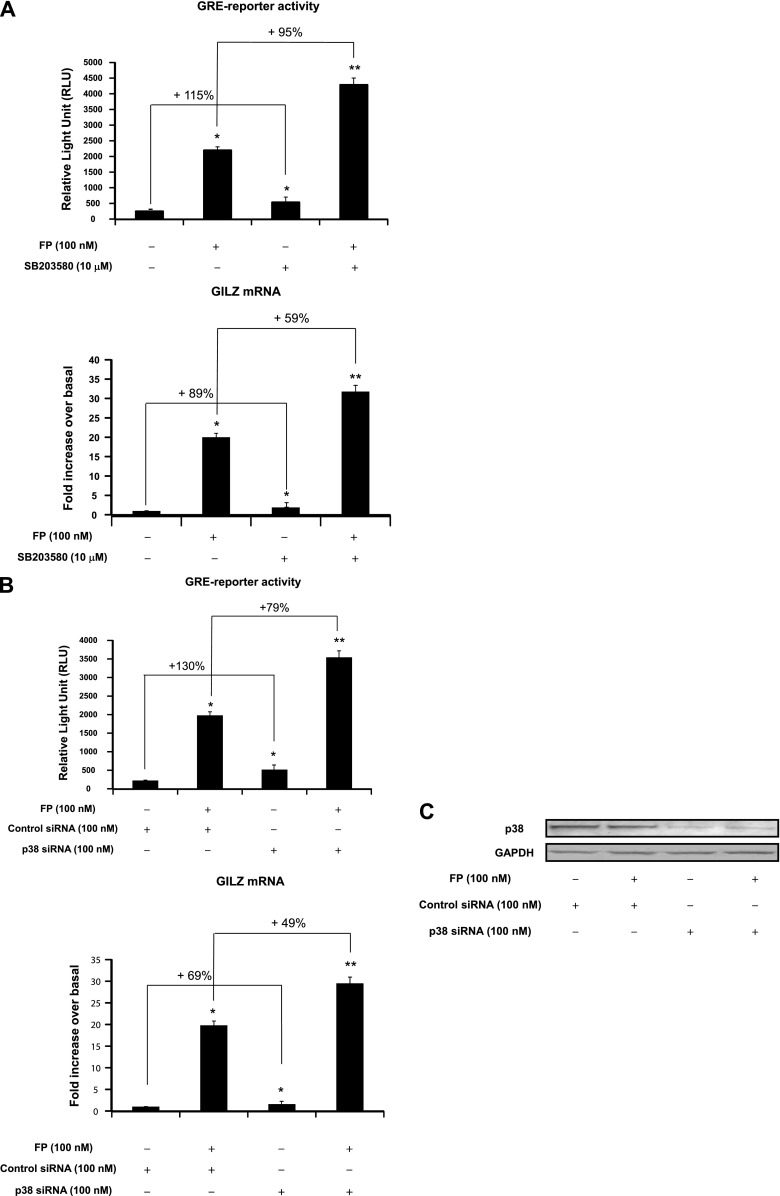
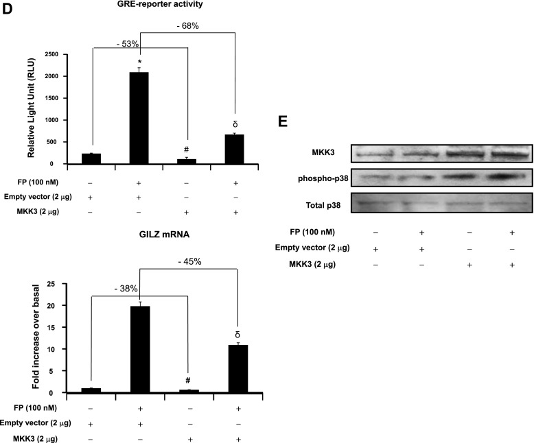
Effect of p38 pathway modulation on GR-mediated transactivation activities. (A, B, and D, top) Cells were transfected for 48 hours with 2 μg luciferase reporter plasmid driven by glucocorticoid-responsive element (GRE) motifs and 1 μg of β-galactosidase vector (A) and/or 100 nM p38 MAPK siRNA or control siRNA (B) and/or 2 μg MKK3 construct or the corresponding empty vector (D). Cells were pretreated with FP alone (100 nM) for 2 hours (A, B, and D) or with SB203580 (10 μM) for 1 hour before FP addition. Cells were lysed, and the luciferase activity was measured as described in Materials and Methods. The results are expressed as relative light unit (RLU). Data are representative of three separate experiments. (A, B, D, bottom) ASM cells were left untransfected (A) or transfected for 48 hours with 100 nM p38 MAPK siRNA or control siRNA (B) and/or 2 μg MKK3 construct or the corresponding empty vector (D). Cells were treated as above. Total mRNA was analyzed by real-time PCR. The values indicate expression of the steroid-inducible gene, glucocorticoid-induced leucine zipper (GILZ), normalized to GAPDH mRNA levels and presented relative to the expression of basal cells, which was set as 1. Data are representative of three separate experiments. *P < 0.05 compared with untreated cells (A) or with siRNA control–transfected cells (B) or with empty vector–transfected cells (D). **P < 0.01 compared with untreated cells (A), with siRNA control-transfected cells (B), or with empty vector–transfected cells (D). #P < 0.05 compared with empty vector–transfected cells left untreated (D). δP < 0.05 compared with empty vector–transfected cells treated with FP alone (D). (C, E) Cells were transfected for 48 hours with 100 nM p38 MAPK siRNA or control siRNA (C) or with 2 μg MKK3 construct or the corresponding empty vector (E) before FP (100 nM) was added for 2 hours. Cells were lysed, and whole cell lysate extracts were prepared and assayed for p38 MAPK and GAPDH (C) or MKK3, phospho-p38 MAPK, and total p38 MAPK (E) by immunoblot analysis. Results are representative of three separate blots.


