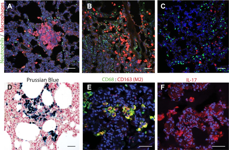
Figure 6.
E. coli sepsis leads to persistent inflammation in the lung. (A–C) Immunostaining for neutrophil elastase (green) and macrophage marker, CD68 (red), showed significant accumulation of interstitial macrophages in foci within the lung parenchyma (A) of an animal killed after 6.5 months (A) and 27 months (B) after experimental sepsis as compared with a healthy control animal (C). The inflammatory infiltrates were frequently associated with the periphery of the organ or with the airway ducts, as shown in (B). Quantification of these data is shown in Figure 1G. Although not statistically significant, neutrophil count was also increased, particularly in the areas surrounding macrophage accumulation. (D) Staining with Prussian blue showed accumulation of iron-positive (blue) hemosiderin-laden macrophages. (E) Costaining for CD68 (green) and CD163 (red; colocalization, yellow) suggests that the macrophages accumulated may belong to the M2 subtype. (F) Immunostaining for IL-17 shows presence of IL-17–positive cells (red) with macrophage morphology. Scale bars, 200 μm (A, B, and D), 100 μm (E and F).

