Figure 1.
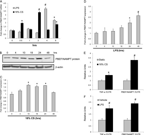
Effects of inflammatory agonists on pre–B-cell colony-enhancing factor (PBEF)/NAMPT expression and 3′-untranslated region (UTR) reporter activity. Total RNA was isolated from human pulmonary artery endothelial cells (ECs) challenged with control vehicle or LPS (100 ng/ml) or exposed to 18% cyclic stretch (CS) for 0 to 8 hours. PBEF/NAMPT mRNA levels were detected via real-time PCR (A). Data are presented as fold change in mRNA level over vehicle-treated control and expressed as mean ± SE from three independent experiments. *P < 0.05 versus unstimulated control. #P < 0.01 versus unstimulated control. Confluent ECs were treated with control vehicle or 18% CS (B) and LPS for the indicated times, and endogenous PBEF was detected via immunoblot. The bar graphs represent relative densitometry (18% CS [C]; LPS [D]). Data are presented as fold changes in PBEF/NAMPT over vehicle-treated control and expressed as means ± SE from three independent experiments. *P < 0.05 versus unstimulated control. #P < 0.01 versus unstimulated control. ECs were cotransfected with PBEF/NAMPT 3′-UTR reporter together with phRL-TK, a Renilla luciferase normalization control vector, and exposed to 18% CS (E) or treated with LPS (F) (24 h), and luciferase activity was measured using the Dual Luciferase Assay System according the manufacturer’s protocol. TNF-α luciferase reporter was used as a positive control. The bar graph represents relative luciferase units. Data are presented as relative luciferase units over vehicle-treated control and expressed as means ± SE from three independent experiments. *P < 0.05 versus unstimulated control. #P < 0.01 versus unstimulated control.

