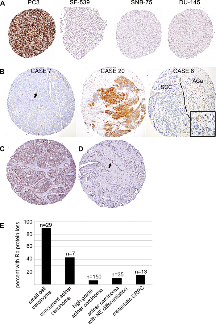
Figure 1.
(A) Immunostaining for Rb was validated using a tissue microarray made from the NCI-60 cell line panel. PC3 cells show high Rb expression with wildtype RB1. SF-539 cells are negative for Rb protein and are known have a 4 base pair deletion resulting in a frameshift mutation. SNB-75 cells are not known to have a homozygous deletion in RB1, however numerous previous studies have shown absence of the Rb protein by western blotting in this cell line. DU-145 cells have a homozygous deletion resulting in a truncation of the protein at exon 21 and show very low Rb staining by immunohistochemistry. (B) Representative Rb immunostaining results from Case 7 shows negative staining in tumor cells with retained endothelial staining (arrow) as an internal positive control. Case 20 shows strongly positive staining. Case 8 shows negative staining in the small cell carcinoma (SCC) component and positive staining in the adjacent acinar carcinoma component (ACa). Images in this panel were assembled from a mosaic of higher power images. (C) Representative positive Rb immunostaining results in a high grade acinar carcinoma unassociated with small cell carcinoma. (D) Representative negative Rb immunostaining in a high grade acinar carcinoma unassociated with small cell carcinoma. Endothelial cells (arrow) provide an internal positive control. (E) Graphical representation of percent of cases with Rb protein loss comparing small cell carcinomas, acinar carcinomas associated with small cell carcinoma and high grade acinar carcinomas unassociated with small cell carcinoma. Rb loss is most frequent in small cell carcinomas and rarely seen in high grade acinar carcinomas unassociated with small cell carcinoma, while acinar carcinomas occurring concurrently with small cell carcinoma show an intermediate rate of Rb protein loss.

