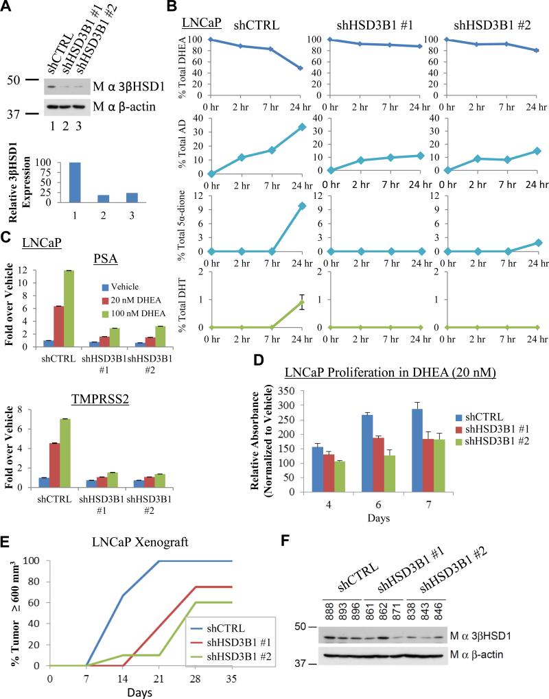
Fig. 3.
Genetic silencing of 3βHSD1(367T) impedes conversion of DHEA to DHT, induction of PSA and TMPRSS2 expression, and CRPC growth. (A) Stable lentiviral expression of two independent shRNA constructs against HSD3B1 (shHSD3B1 #1 and shHSD3B1 #2) silences 3βHSD1 protein expression in LNCaP. The 3βHSD1 protein was quantitated and normalized to cells expressing nonsilencing lentiviral vector (shCTRL) and β-actin. (B) Silencing 3βHSD1(367T) blocks flux from [3H]-DHEA (100 nM) to AD as well as further downstream conversion to 5α-dione and DHT. Cells were treated with [3H]-DHEA in triplicate and steroids were quantitated with HPLC at the designated time points. (C) Inhibition of AR-regulated genes. Cells were treated with the indicated concentration of DHEA for 24 hours, and gene expression was assessed by qPCR and normalized to shCTRL-infected cells treated with vehicle and the RPLP0 housekeeping gene. (D) Silencing 3βHSD1(367T) inhibits in vitro growth. Cells were grown in the presence of 20 nM DHEA or vehicle and growth for each cell line is normalized to vehicle for each designated day. (E) 3βHSD1(367T) depletion blocks CRPC growth in LNCaP xenografts. Mice underwent surgical orchiectomy and DHEA pellet implantation concomitantly when xenograft tumors reached a threshold volume of 100 mm3. Fifteen mice were initiated in each cohort, 7, 8 and 10 mice in shCTRL, shHSD3B1 #1, and shHSD3B1 #2 groups, respectively, achieved a tumor volume of 100 mm3 in eugonadal mice, underwent orchiectomy and were included in the CRPC analysis. The number of days from orchiectomy to tumor volume ≥ 600 mm3 is shown. In the comparisons of shCTRL vs shHSD3B1 #1 and shHSD3B1 #2, P = 0.002 and 0.003, respectively, using a log rank test. (F) 3βHSD1(367T) protein is regained in CRPC tumors that grow from LNCaP expressing shHSD3B1 #1 and shHSD3B1 #2. Immunoblot for 3βHSD1 and β-actin were performed on protein from the indicated LNCaP CRPC tumors. Error bars in B, C and D represent the SD for experiments performed in triplicate.

