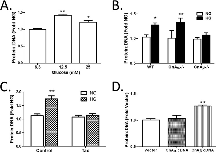
FIGURE 2.
CnAβ mediates cellular hypertrophy. A, hypertrophy was examined in WT kidney fibroblasts exposed to increasing amounts of glucose for 48 h by assessing the protein/DNA ratio. Data shown are the mean ± S.E. (error bars) of three independent experiments. *, p < 0.05; **, p < 0.01 compared with low glucose. B, hypertrophy was examined in WT, CnAα−/−, or CnAβ−/− kidney fibroblasts treated with NG or HG for 48 h by assessing the protein/DNA ratio. Data shown are the mean ± S.E. of three independent experiments relative to WT NG. *, p < 0.05; **, p < 0.01. C, hypertrophy was assessed in tacrolimus-treated (Tac) CnAα−/− kidney fibroblasts exposed to NG or HG for 48 h. Data shown are the mean ± S.E. of 8 replicates/group relative to control NG. **, p < 0.01. D, hypertrophy was assessed in vector, CnAα, or CnAβ cDNA-transfected WT kidney fibroblasts after 72 h of expression. Data shown are the mean ± S.E. of three independent experiments relative to vector. **, p < 0.01.

