FIGURE 5.
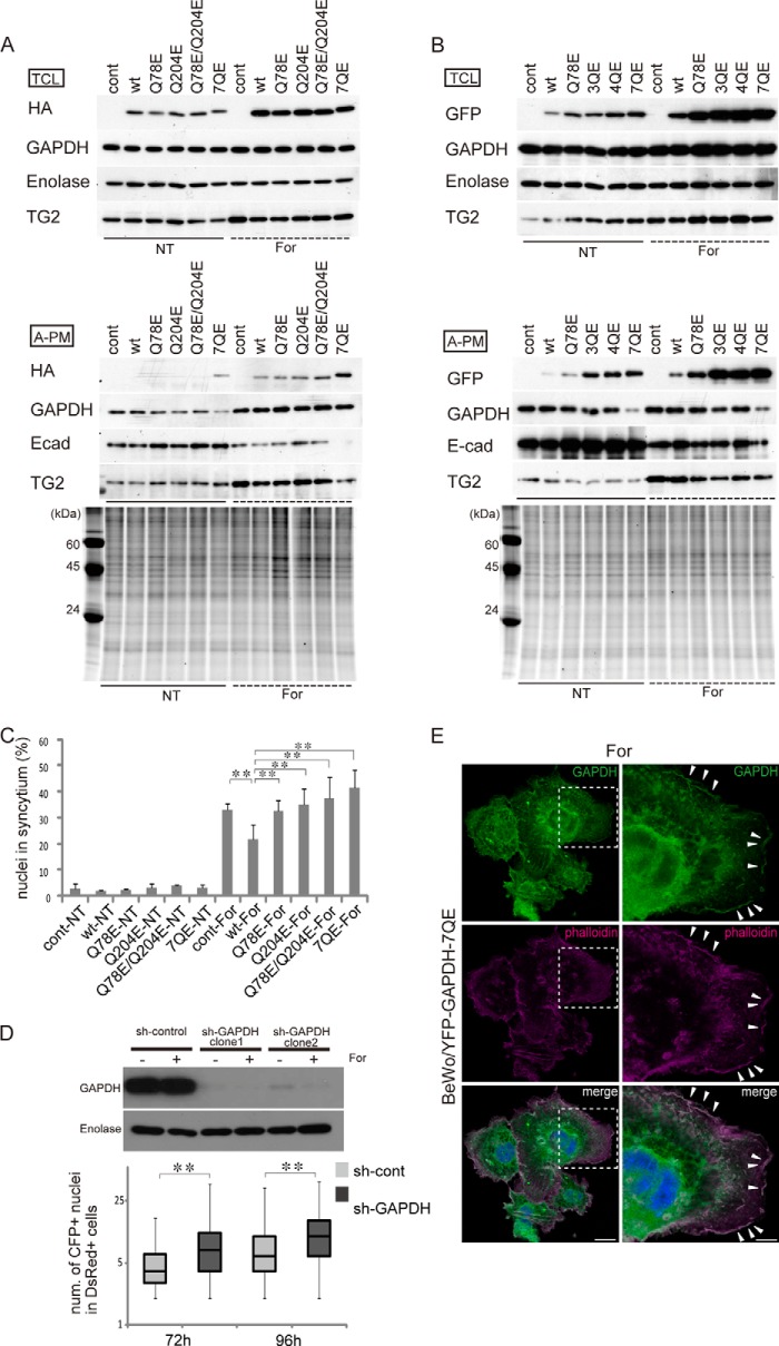
Effects of Gln/Glu-substituted GAPDH mutants on cell fusion. A, Western blots of wild-type and HA-tagged mutant GAPDH in total cell lysate (TCL) and the A-PM. HA-tagged wild-type, Q78E, Q204E, Q78E/Q204E, or 7QE mutated GAPDH was expressed in BeWo cells using a lentivirus vector. The cells were cultured in the presence or absence of 25 μm forskolin for 72 h. All of the Gln residues were replaced by Glu in the 7QE mutant. For A-PM samples, the SYPRO Ruby protein stain images of the SDS-polyacrylamide gel are presented in the bottom panel. The endogenous GAPDH and HA-tagged mutant were not discriminated from each other in the GAPDH band. B, Western blots of wild-type and EYFP-tagged GAPDH mutants. EYFP-tagged wild-type, Q78E, Q204E/Q264E/Q280E (3QE), Q48E/Q78E/Q113E/Q185E (4QE), or 7QE GAPDH was expressed in BeWo cells using a lentivirus vector. The cells were cultured in the presence or absence of 25 μm forskolin for 72 h. For A-PM samples, the SYPRO Ruby protein stain images of the SDS-polyacrylamide gel are presented in the bottom panel. The GAPDH band does not include EYFP-tagged fusion GAPDH. C, percentage of syncytial cell nuclei among total nuclei in the HA-tagged experiment. More than 100 nuclei in each microscopic field (×100 magnification) were counted (n = 5). Error bars, S.D. **, p < 0.01 (Tukey-Kramer statistical test). D, effects of GAPDH knockdown on cell fusion. Western blot of GAPDH in the total cell lysate of BeWo cells expressing scrambled shRNA (sh-control) or shRNA of GAPDH (sh-GAPDH) (top). The cells were cultured in the presence or absence of 25 μm forskolin for 72 h. Bottom, numbers of nuclei in the fused cells 72 and 96 h after forskolin treatment. Two different cells harboring either red fluorescent protein in the cytoplasm (DsRed cells) or enhanced cyan fluorescent protein with a nuclear localization signal (CFP-Nuc cells) were co-cultured, and nuclei were counted in the double positive cells (n > 300). E, localization of the YFP-GAPDH-7QE mutant. The mutant GAPDH co-localizes with F-actin at the plasma membrane. Scale bars, 25 μm (left) and 10 μm (right). The fluorescent image was obtained with a Leica TCS SP8 confocal fluorescent microscope.

