FIGURE 2.
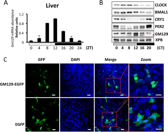
GM129 is a nuclear protein and oscillates in mouse liver. A, circadian oscillation of Gm129 mRNA in mouse liver. Mice were sacrificed at zeitgeber times (ZT) 4, 8, 12, 16, 20, and 24/0 h. The abundance of Gm129 mRNA was determined by quantitative RT-PCR and normalized to that of Gapdh. The maximal value was normalized to 1. Error bars represent S.D. of 2 biological repeats. The 0 to 24-h time point is plotted twice to emphasize the trend. B, oscillation of GM129 and core clock protein levels in liver nuclei. Mice were kept in constant dark for 24 h following 12-h dark/12-h light cycles. After 24 h in the dark, nuclear extracts were prepared from livers collected at 0, 4, 8, 12, 16, and 20 h in dim red light and GM129 was detected by Western blots. The XPB protein was used as a loading control. The GM129 signal is marked by a red asterisk. Note there is a nonspecific antibody cross-reactive band above the GM129 signal. The blot is representative of two experiments. C, subcellular localization of GM129-EGFP fusion protein. HEK293T cells were transfected with plasmid DNA expressing GM129-EGFP protein or EGFP alone. 48 h post-transfection, cells were fixed and visualized by fluorescence microscopy (GFP) and DAPI staining. Nuclear localization was confirmed by merging the GFP signal with the DAPI signal. EGFP alone is a control that localizes to both cytoplasm and nucleus. Bar = 10 μm.

