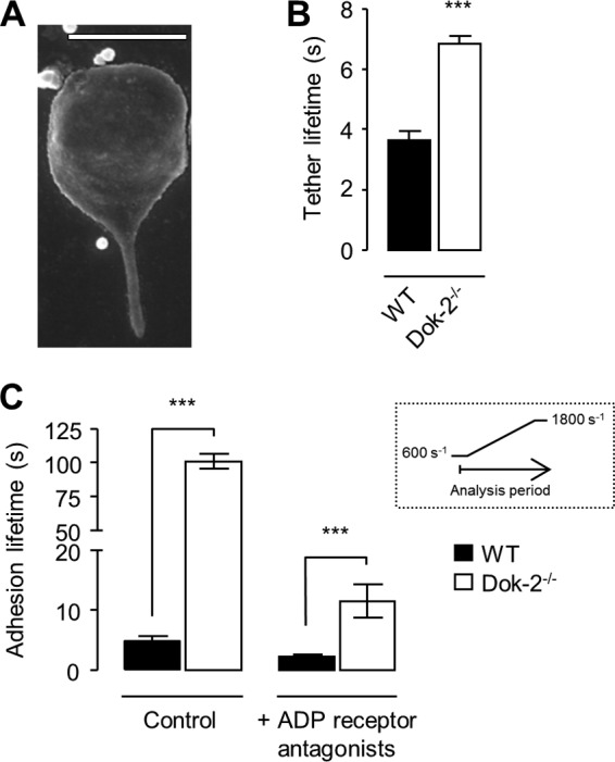
FIGURE 5.

Dok-2 deficiency leads to an increased adhesion lifetime of platelet membrane tethers. Anticoagulated whole blood from WT or Dok-2−/− mice was perfused over fibrinogen (20 μg/ml)-coated microslides at 600 s−1. A, a cropped SEM image of a representative murine platelet tether (WT). The image was taken from one experiment representative of three. Scale bar = 2 μm. B, tether adhesion lifetime was determined by counting the number of frames for which a platelet was tethered to the matrix, as described under “Experimental Procedures.” ***, p < 0.001. C, whole blood was perfused over fibrinogen in the presence or absence of ADP receptor antagonists (MRS2179 (400 μm) and 2-methylthioadenosine 5′-monophosphate triethylammonium salt (2MeSAMP) (40 μm)) and apyrase (0.02 units/ml) at 600 s−1 for 2 min. The shear rate was increased to 1800 s−1 for 3 min. The histogram shows the time (s) platelets remained attached (adhesion time) during the shear rate increase as a measure of bond strength. The inset shows a schematic of the analysis period. Data represent the mean ± S.E., n = 3. ***, p < 0.001.
