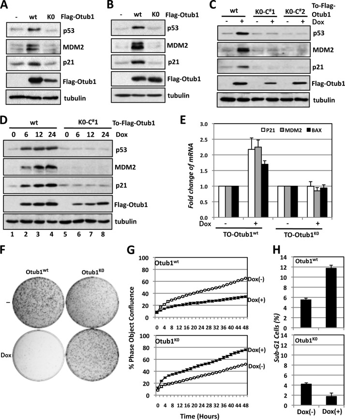
FIGURE 3.
Lysine-free Otub1 fails to induce and activate p53. A and B, Otub1K0 does not induce p53. U2OS cells were transfected with equal amounts (3 μg) of the FLAG-Otub1wt or FLAG-Otub1K0 plasmid (A) or 0.5 μg of the FLAG-Otub1wt or 3 μg of the FLAG-Otub1K0 plasmid (B), followed by IB analysis. C, induced expression of Otub1K0 fails to induce p53. Two representative U2OS-TO-FLAG-Otub1K0 clones and the U2OS-TO-FLAG-Otub1wt stable cell line were cultured in the absence or presence of 2 μg/ml of Dox for 24 h. The cell lysates were assayed by IB analysis. D, time course study of the p53 induction by Otub1wt but not Otub1K0. U2OS-TO-FLAG-Otub1wt or U2OS-TO-FLAG-Otub1K0 clone 1 (C#1) was cultured in the presence of 2 μg/ml of Dox and harvested at different time points, as indicated, followed by IB analysis. E, Otub1K0 does not induce p53 activity. U2OS-TO-FLAG-Otub1K0 or U2OS-TO-FLAG-Otub1wt cells were treated with or without Dox for 24 h, followed by RT-qPCR detection of p21, mdm2, and bax mRNA normalized to the expression of GAPDH. F, Otub1K0 does not inhibit cell proliferation. U2OS-TO-FLAG-Otub1wt or U2OS-TO-FLAG-Otub1K0 cells were cultured in the presence or absence of Dox for up to 3 weeks. The colonies were visualized by staining with crystal violet blue. G, T-Rex-U2OS-FLAG-Otub1wt or T-Rex-U2OS-FLAG-Otub1K0 cells were cultured in the presence or absence of 2 μg/ml doxycycline. The cell confluence was measured over time using the IncuCyte system. H, Otub1K0 does not induce apoptosis. U2OS-TO-FLAG-Otub1wt or U2OS-TO-FLAG-Otub1K0 cells were cultured with or without Dox for 24 h. The cells were stained with propidium iodide, followed by flow cytometry analysis. The average percentages of cells in sub-G1 are shown.

