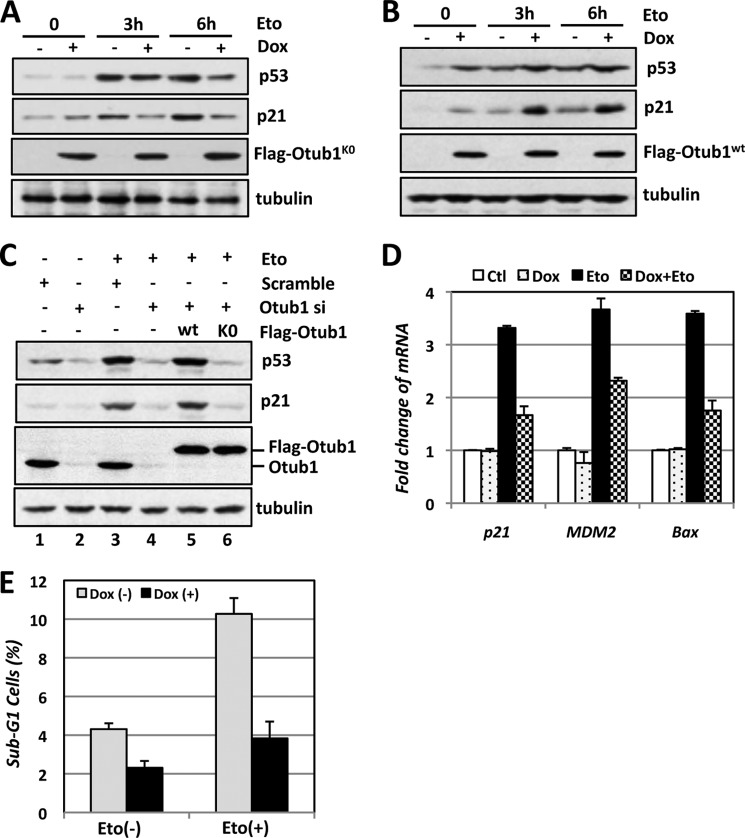
FIGURE 4.
Lysine-free Otub1 attenuates p53 activation in response to DNA damage. A and B, Otub1K0, but not Otub1wt, attenuates p53 activation following Eto treatment. U2OS-TO-FLAG-Otub1K0 cells (A) and U2OS-TO-FLAG-Otub1wt cells (B) were cultured in the absence or presence of Dox for 24 h, followed by treatment with Eto (20 μm) or control dimethyl sulfoxide for the indicated times, followed by IB analysis. C, reintroduction of Otub1wt, but not the Otub1K0 mutant, rescues the p53 response following DNA damage in cells with Otub1 knockdown. U2OS cells transfected with control, the FLAG-Otub1wt or FLAG-Otub1K0 plasmid, and scrambled or Otub1 siRNA targeting the 3′ UTR of the Otub1 mRNA are shown, as indicated. The cells were treated with Eto (20 μm) for 6 h before harvesting 48 h after siRNA transfection, followed by IB analysis. D and E, Otub1K0 attenuates p53 activation in response to DNA damage. U2OS-TO-FLAG-Otub1K0 cells were cultured in the absence or presence of Dox for 24 h, followed by treatment with Eto (20 μm) or control (Ctl) dimethyl sulfoxide for 6 h, and assayed by RT-qPCR (D) and cell cycle profile analysis for sub-G1 phase cells (E).

