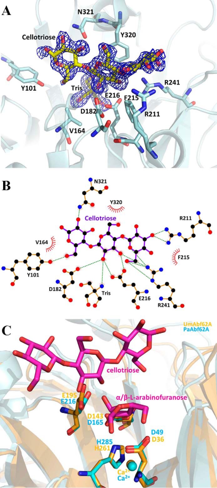
FIGURE 7.

PaAbf62A in complex with cellotriose. A, structure of cellotriose in the active site pocket of PaAbf62A. Sugar is displayed as stick and protein as a schematic. Catalytic amino acids are named using the one-letter code of the amino acid followed by the position in the sequence. The electron density is displayed in dark blue and has been contoured to 1σ. B, schematic diagram showing hydrogen-bonding, water-bridged, and hydrophobic interactions between PaAbf62A and cellotriose. Amino acid residues of PaAbf62A that have hydrophobic interactions with cellotriose are shown as spiked spheres (with distances of less than 3.5 Å). Direct and water-bridged hydrogen-bonding interactions are indicated by dashed lines. C, superimposition of complex structures of PaAbf62A and UmAbf62A sugar are colored in magenta, UmAbf62A is colored in gold, and PaAbf62A is colored in cyan.
