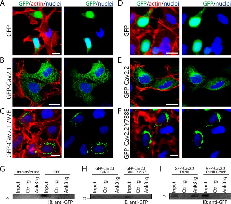
FIGURE 8.
Cav2.1/Cav2.2 tyrosine residues in ABM are critical for cellular targeting. HEK293 cells transfected with pEGFP-C3 demonstrated a primarily nuclear localization pattern (A and D), whereas HEK293 cells transfected with Cav2.1 DII/III-GFP (B) or Cav2.2 DII/III-GFP (E) displayed a diffuse cellular pattern of expression unique from the nucleus. C and F, unlike WT constructs, both Cav2.1 Y797E-GFP and Cav2.2 Y788E-GFP were distributed in a tight perinuclear pattern. Scale bars denote 8 μm. G–I, a conserved tyrosine residue in the Cav2.1 and Cav2.2 DII/III loop is necessary for ankyrin-B interaction in vitro. G, in control experiments, ankyrin-B Ig did not interact with GFP alone expressed in HEK293 cells. H and I, ankyrin-B Ig co-immunoprecipitates GFP fusion protein containing the Cav2.1 or Cav2.2 DII/DII loop from transfected HEK293 cells. In contrast, ankyrin-B Ig does not co-immunoprecipitate GFP fusion proteins of Cav2.1 DII/DIII Y797E or Cav2.2 DII/III Y788E in parallel experiments. IB, immunoblotting.

