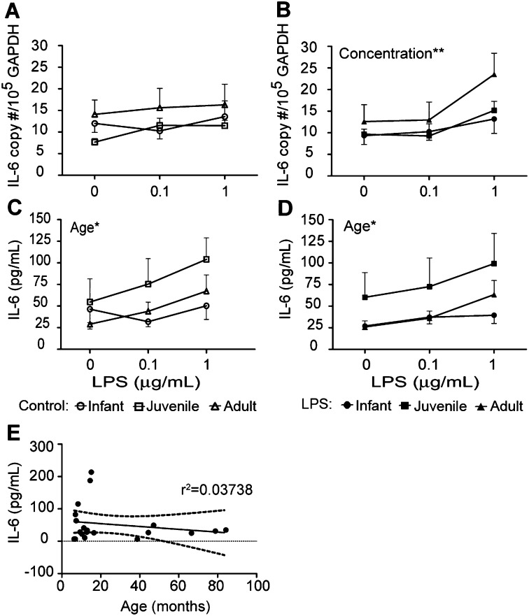
Figure 4.
Effects of age and previous exposure on LPS-induced IL-6 expression in airway epithelium. Tracheal slices were cultured with LPS for 24 hours, followed by protease digestion and the isolation of airway epithelial cells. Air–liquid interface cultures from infant, juvenile, or adult monkeys received a second treatment of LPS (0.1–1 μg/ml), and were evaluated for IL-6 expression after 24 hours. (A and B) IL-6 mRNA and (C and D) IL-6 protein expression was determined in cultures established from media control (A and C) and LPS-exposed (B and D) tracheal slices. Each data point represents the mean ± SE from 5–6 animals per group. *P < 0.05 and **P < 0.01, according to two-way ANOVA, age versus LPS concentration. Circles, infants; squares, juveniles; triangles, adults. Open symbols indicate control cultures, whereas solid symbols indicate ex vivo LPS cultures. (E) Correlation between chronological age (in months) and IL-6 protein secretion in primary airway epithelial cell cultures. Each data point represents basal IL-6 protein concentration (no ex vivo or secondary LPS) in airway epithelial cell cultures derived from individual animals.

