Figure 1.
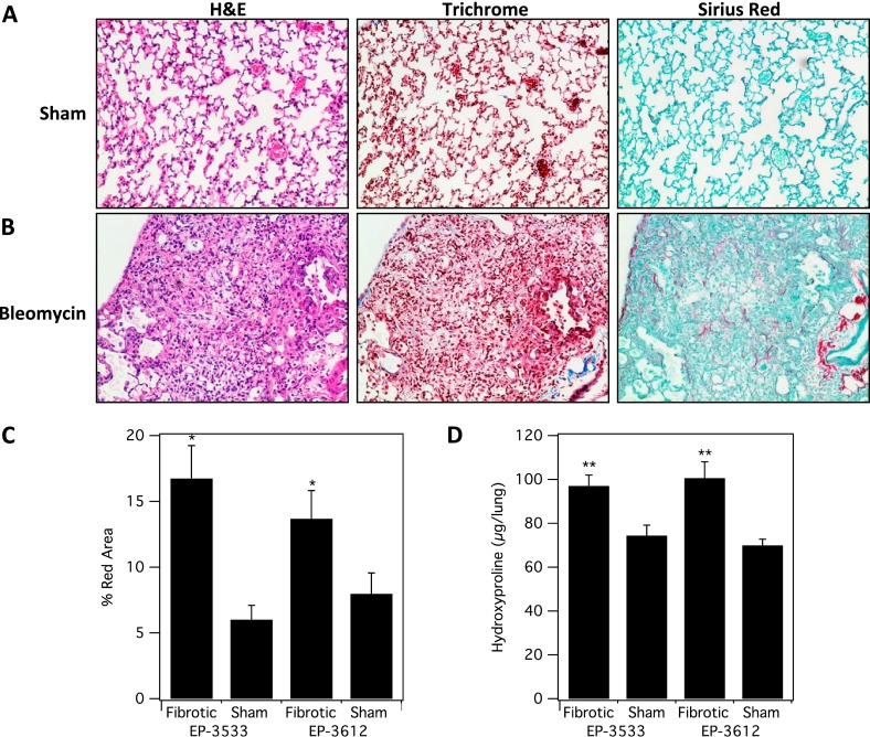
Characterization of fibrosis and collagen deposition in the mouse bleomycin (BM) model. Representative images of lung tissue stained with hematoxylin and eosin (H&E), trichrome, and Sirius red for sham (A) and BM-treated mice (B). (C) Sirius red staining was quantified and was significantly higher in fibrotic (BM-treated) mice (*P < 0.001). (D) Hydroxyproline (Hyp) analysis of ex vivo harvested lung tissue from all four cohorts shows significantly higher Hyp/lung in the fibrotic (BM) -treated cohorts (**P < 0.0001).

