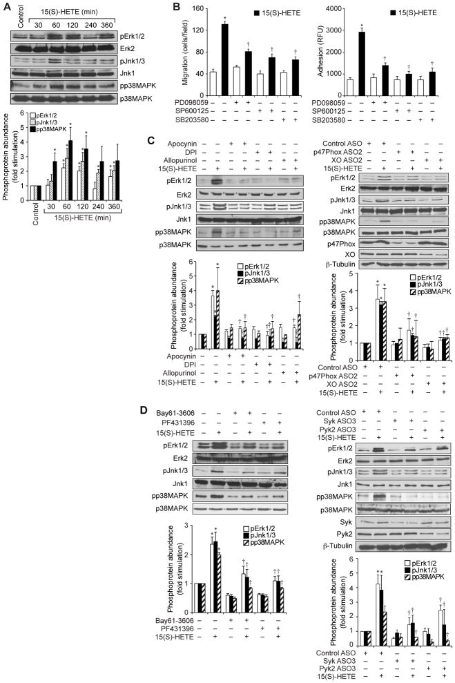
Fig 5. MAPKs mediate the 15(S)-HETE–dependent migration and adhesion of THP1 cells.
(A) Whole-cell lysates of vehicle-treated THP1 cells (Control) or of THP1 cells treated with 0.1 μM 15(S)-HETE for the indicated times were analyzed by Western blotting with antibodies specific for the indicated proteins. Bar graph shows densitometric analysis of the fold-increase in the abundance of pErk1/2, pJnk1/3 and pp38MAPK at the indicated times of 15(S)-HETE treatment compared to that in vehicle-treated cells. (B) THP1 cells were pretreated for 30 min with vehicle, 30 μM PD098059, 10 μM SP600125, or 10 μM SB203580 before being treated with vehicle or 0.1 μM 15(S)-HETE and then being analyzed for migration (left) and adhesion (right). (C and D) THP1 cells were treated with the indicated inhibitors or were transfected with the indicated ASOs before being treated with vehicle or 0.1 μM 15(S)-HETE for 1 hour. Whole-cell lysates were then analyzed by Western blotting with antibodies specific for the total and phosphorylated forms of the indicated MAPK proteins, p47Phox, XO, Syk, and Pyk2. β-tubulin was used as a loading control. Bar graph shows densitometric analysis of the fold-increase in the abundance of pErk1/2, pJnk1/3 and pp38MAPK for each condition compared to that in vehicle-treated cells. Western blots are from one experiment and are representative of three independent experiments. Data in bar graphs are means ± SD from three independent experiments. *P < 0.01 versus control; †P < 0.01 versus 15(S)-HETE.

