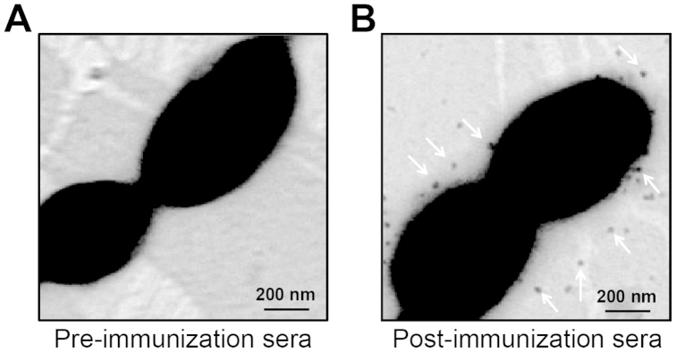
Figure 5. Immune electron microscopy-based analyses of the BgaC protein.

The BgaC protein is visulized to display on bacterial cell surface of S. suis 2 using the post-immunization sera (Panel B), whereas not using the pre-immunization sera (Panel A).
