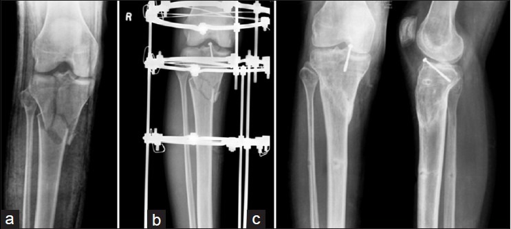
Figure 4.

(a) X-ray anteroposterior view of knee joint showing tibial plateau fracture (b) x-ray anteroposterior view treated with minimal internal fixation and a ring fixator (c) X-ray anteroposterior and lateral views showing negligible plateau tilt and articular step
