
Figure 2.
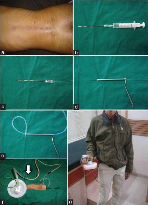
Procedure Illustration (a) The anatomical clinical surface markings with the entry point for the abscess marked patient in prone position. (b) The epidural needle is inserted to a depth less by 2 cm calculated already, then stylet is withdrawn and syringe aspiration done. (c) A 0.5 mm long guide wire is threaded into the needle. (d) The needle is withdrawn and then over the guide wire the serrated biopsy cannula is plunged over the guide wire. (e) The guide wire is withdrawn and the catheter is threaded through the trocar and trocar is then withdrawn. (f) Draining pus flow (arrow). (g) Patient is ambulatory with the PCD