
Figure 3.
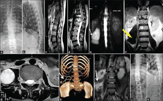
(a and b) Anteroposterior and lateral radiograph showing D11-12 paradiscal affection. (c and d) MRI sagittal T1W and T2W showing D11-D12-L1 spondylodiscitis. (e and f) MR Myelogram and T1 coronal section showing the huge abscess extending from L1 to L5, with maximum width at L3 lower border in the right side (yellow arrow). (g) MRI T2 axial image at L 3 lower border. The estimation from this section is used to decide the desired point of aspiration by our technique (details in Figure 1). (h) 3D CT reconstruction image showing the coiled catheter in the abscess. (i) MRI T 2 coronal image showing the collapsed aspirated abscess cavity (white arrow). (j and k) Anteroposterior and lateral radiograph showing healed sclerosed vertebrae at 18 months