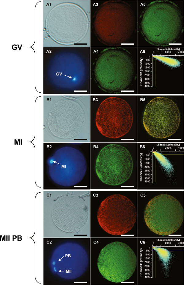
Figure 2.
Confocal images of mt distribution, ROS levels and mt/ROS colocalization of dromedary camel oocytes. Mitochondrial distribution patterns and intracellular reactive oxygen species (ROS) localization in dromedary camel oocytes at GV (Panel A), MI (Panel B), and MII (Panel C) stage observed after staining with MitoTracker Orange CMTM Ros, H2DCF-DA and Hoechst 33258. An immature GV stage oocyte showing homogeneous mt distribution pattern of small mitochondrial aggregates (Panel A), a MI oocyte showing heterogeneous distribution of mitochondria (pericortical tubular network, PCTN; Panel B), and a MII oocyte showing heterogeneous distribution of mitochondria (perinuclear/pericortical tubular network, PN/PCTN; Panel C) are shown. For each sample, corresponding bright field (A1, B1, C1), UV light (A2, B2, C2) and confocal images showing mitochondrial distribution pattern (A3, B3, C3) intracellular ROS localization (A4, B4, C4), mitochondrial/ROS merge (A5, B5, C5) and the mitochondrial/ROS colocalization scatter plot (A6, B6, C6) are shown. The scale bar represents 60 μm.

