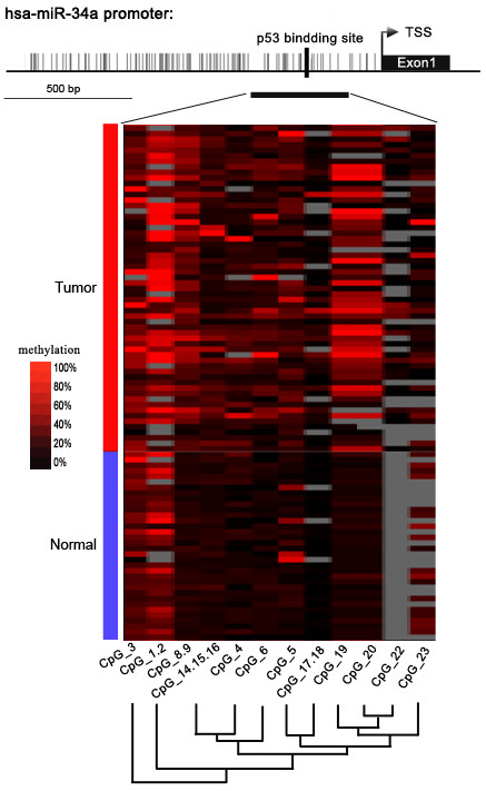
Figure 1.

Genomic structure of distribution of miR-34a CpG dinucleotides over transcription start site (TSS) and hierarchical cluster analysis of CpG units’ methylation profiles of miR-34a promoter region in tumor (n = 59) and normal (n = 34) tissues. The depicted region corresponds to 1.2 kbp upstream of the TSS (indicated by arrow). Each vertex indicates an individual CpG site. The positions and orientation of the MassARRAY primers are indicated by horizontal black bars. The position of the p53 binding site is indicated. Columns display the clustering of CpG units, which are a single CpG site or a combination of CpG sites. Each row represents a sample. The methylation intensity of each miR-34a CpG unit in each sample varies from red to black, which represents high to low expression. The color gradient between black and red indicates methylation ranging from 0 to 100. Gray represents technically inadequate or missing data.
