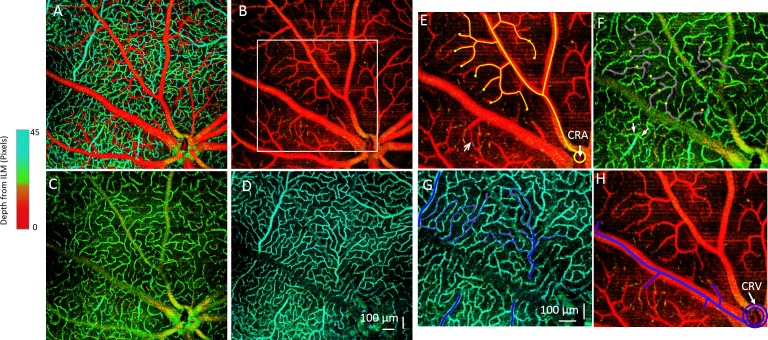
Figure 4.
Depth-resolved retinal microvascular network and 3D tracking of the microvessels are shown. (A) Depth-coded color angiography of the whole mouse retina is shown. (B–D) Microvascular networks are shown within 3 different layers: (B) GCL, (C) IPL, and (D) OPL. The microvascular organization within the 3 layers has distinct patterns. (E–H) Tracking of the microvessel paths of an arteriole and venule is demarcated in the white rectangular region in (B). (E) In the superficial microvessels of the GCL, the blood flows through arterioles branched from the central retinal artery (CRA), shown as the yellow lines. The yellow dots denote where the vessels dive to a separate layer. The arrow points to a direct arteriovenular connection in the superficial GCL layer. (F) An intermediate capillary layer in the IPL is shown. Yellow dots indicate the vessels that are diving to this layer from the superficial layer in (E). These capillaries flow as pink lines and then dive into a deeper layer at the pink dots. Arrows point to capillaries joining the postcapillary venules. (G) Deep-layer microvessel in OPL is shown, in which the pink dots from IPL indicate diving vessels and blue lines indicate flow. All of the blood flow will then be collected by the postcapillary venules and returned to the superficial layer and to the major venules (H), which will join the central retinal vein (CRV).

